Featured Applications
Precision Optics:
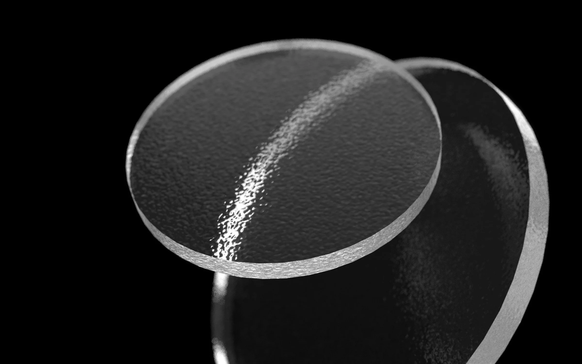
Scratch, Dig, Transmission
MODI can successfully measure nearly any optical component variety—flats, concave mirrors, transparent lenses, infrared lenses, high curvature surfaces, large optics~ 0275mm. We have qualified with standards to MIL 5/5. Spectral transmission can also be measured with the onboard spectrometer.
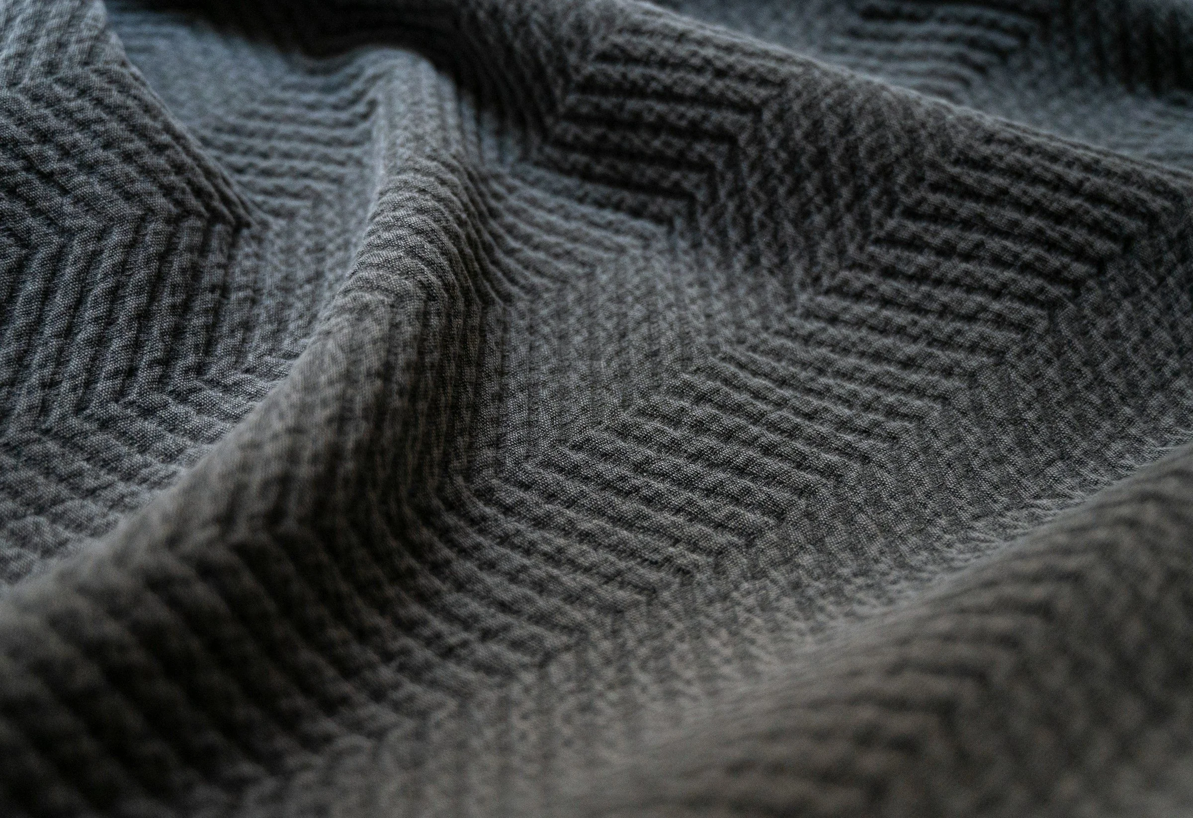
Polymers and Textiles
The MODI system is capable of spectrally fingerprinting and distinguishing between various class of materials, including different blends and composition ratios.

Water Content Imaging
Infrared Wavelengths, particularly bands around 1225nm and 1425nm, exhibit O-H absorption. Water content can be measured and imaged across a sample. Geopulse Solutions has validated leaf water content against traditional mass dehydration methods with a correlation of greater than or equal to 90%.
Coatings and Layer Thickness
Thin film interference patterns measured with a spectrometer are often used to measure coating layer thickness to within nanometers of precision, and track variations.
OPTICS INSPECTION CAPABILITIES
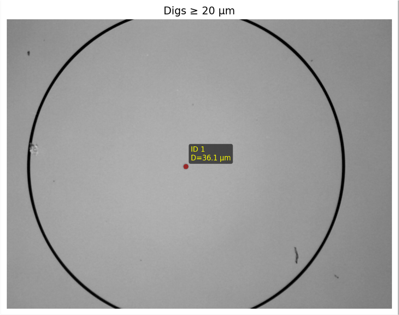
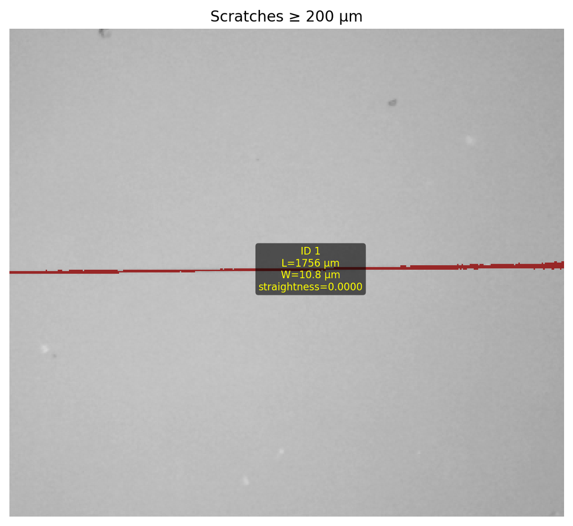
(1) Overlay showing detected and classified surface digs, including labeled defect information such as diameter. (2) Overlay showing detected and classified surface scratches, including labeled defect information such as length & width.
Optics Measurement Standards
MODI is a cost effective solution that meets lab quality measurement standards
MODI has specialized workflows for inspection of precision optical components, allowing for automatic, traceable quality control.
Our automated microscopy solution has been developed for compatibility with a wide variety of optics including lenses, mirrors, and lens assemblies, including larger optics. The solution is resilient to curvature and has been testing on a variety of challenging components. Several device features are critical to this measurement including auto-focus, illumination & smart lighting, and automated defect detection & measurement software.
MODI has certified performance against standards for MIL-PRF-13830B, measuring down to scratch & dig of 5/5 and lower.
Microscopy Features
Digital microscopy goes beyond traditional visual inspection by capturing high-resolution images at the microscale—without eyepieces or manual documentation. It transforms fine detail into quantitative, analyzable data that can be measured, shared, and integrated into automated or AI-driven workflows. This approach reveals structure, defects, and texture while enabling precise measurement of size, shape, and spatial distribution.
Illumination & Smart Lighting
The standard MODI system comes with a variety of integrated lighting types and controls including ring lights, quandrant ring selection, coaxial lighting, darkfield illumination, fluorescence illumination, backlight sources, and scatter light mode.
MODI has scan procedures that auto-detect glare and gradients, adjusting lighting to eliminate any bad data.
Auto-scanning & Mosaicing
Automatic scan routines with our digital microscope are used to raster across a sample and autofocus for each image. The result is built into a mosaic "superimage" that stitches images across the part into one large image, providing high resolution large field pictures.
Focus Stacking
MODI scanning has a critical modality called "focus stacking" that allows a series of hundreds of photos to be taken at different depths, combining all images into a high quality fully focused image. This is particularly important for samples with heavy topology changes.
Automated Defect Detection
Our suite of analysis software tools can be used to detect defects and measure features based on customizable morpholgy and spectral filters. This can be used for example to detect and distinguish Foreign Object Debris (FOO), surface abrasion, scratches, and contamination.
Depth Mapping
Along with focus stacking procedures, you can generate depth maps accurate to the Depth of Field (DoF) of the on board digital microscope. Our Standard configuration microscope has a depth of 0.1mm, allowing depth maps of l00um in accuracy to be generated.
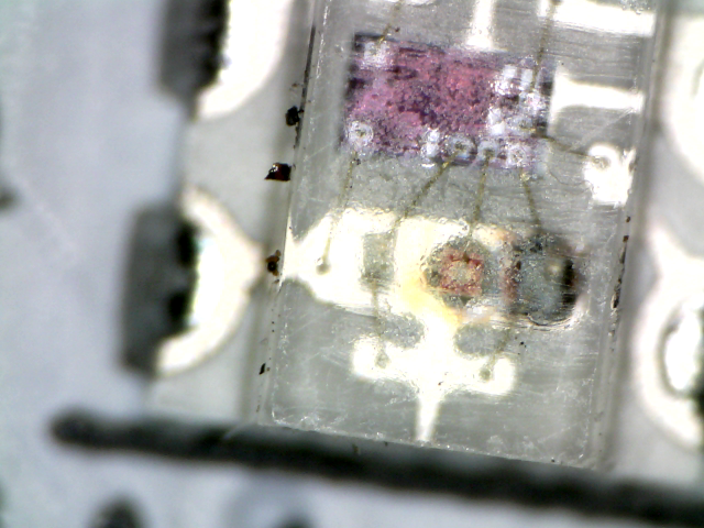
(1) Image from microelectronics failure analysis study (2) Axial light microscopy examining ink from a dry erase marker
Spectral Imaging Features
Imaging spectroscopy extends beyond conventional cameras by capturing the unique spectral “fingerprint” of materials. It produces spatially resolved chemical maps, showing not just what something looks like, but what it is and how much is present. This enables non-destructive identification and quantification of materials, chemicals, and biological states, including their composition, concentration, and distribution.
-
Our Standard configuration includes a Multispectral Module (#1) sensitive to infrared wavelengths. This point sensor is paired with low-noise, high-speed electronics to deliver high quality data. The point is magnified with optics (#2) to provide spatially resolved measurements. Combined with our motion gantry and autofocus, an image is rastered across a sample. This principle applies to the Spectroscopy Plus configuration, using point spectrometers and generate hyperspectral imagery.
-
The Optical Modifier Tool Exchange uses magnetic coupling to pick up and remove different filters, lenses, diffusers, and other optical modifiers from the filter bay (#3). With bandpass filters, the spectral module can be used for quantitative applications that require discrete multispectral sensing.
-
To maintain high quality and consistent results, MODI includes onboard calibration targets that provide known reflectance & emissivity. Calibration is automatically measured and maintained to compensate for any dark current drift, thermal drift, and changes in sensitivity.
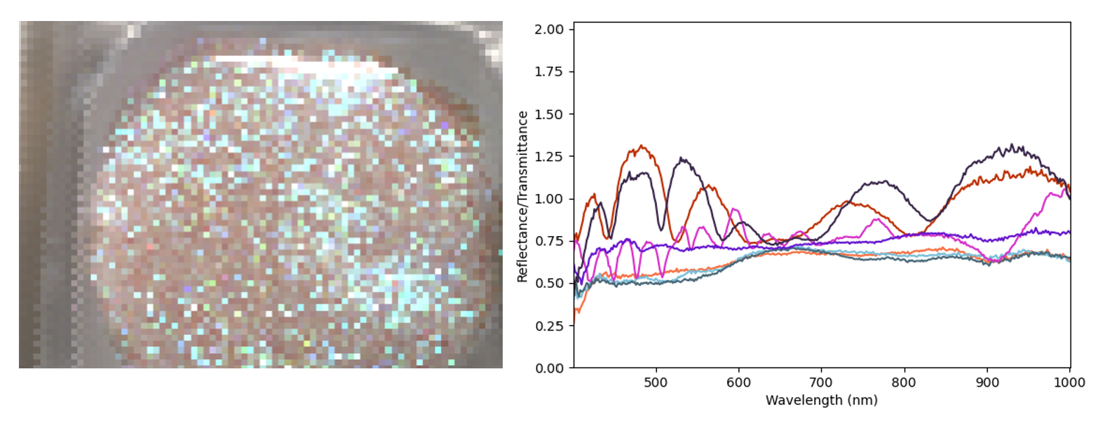
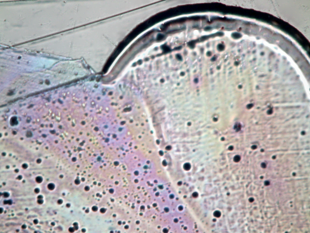
(1) Hyperspectral Image of Mica Flakes in Epoxy (2) Representative Spectral Signatures demonstrating iridescence