Automating Microplastic Research with MODI
RETHINKING MICROPLASTIC ANALYSIS AND WORKFLOW
The MODI platform addresses common microplastic analysis challenges through automation, repeatability, and multimodal data collection.
Microplastics have become one of the most widely studied environmental contaminants, appearing in oceans, rivers, soils, drinking water, food products, and even the atmosphere. As research efforts expand, scientists face a common challenge: collecting large volumes of high-quality, repeatable data efficiently.
Traditional microplastic analysis often requires significant manual effort, or costly lab testing. Researchers may spend hours preparing samples, capturing microscope images, identifying particles, measuring morphology, and documenting results. These workflows can limit throughput and introduce variability between operators and laboratories.
The MODI (Modular Object Detection and Imaging) platform from GeoPulse Solutions was developed to address these challenges through automation, repeatability, and multimodal data collection.
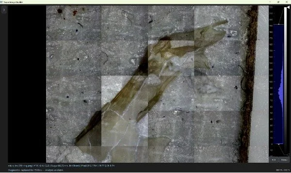
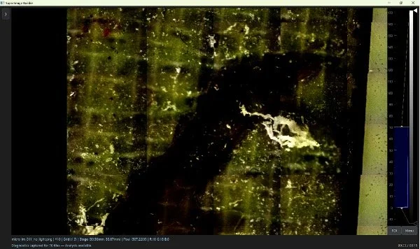
Figure 1 (two images)—Extreme Darkfield illumination technique reveals hidden microplastics on sample surface. Average particle size is measured at ~40um.
MODI combines automated microscopy, image stitching, focus stacking, and advanced illumination techniques to rapidly document entire filters or sample substrates at high resolution. Researchers can automatically acquire large-area datasets while maintaining um-level resolution and consistent imaging conditions across studies and time periods.
Beyond traditional microscopy, MODI supports multispectral and hyperspectral sensing capabilities that can provide additional information about particle composition. Our higher-resolution spectroscopy modalities can reach 50um in spatial resolution, allowing for classification of microscopic particles. By combining morphology, color, spectral response, and spatial context, researchers can develop more robust workflows for particle classification and screening.
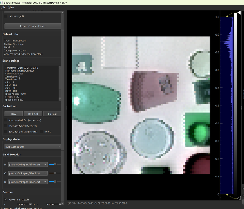
Figure 2—Microscopic Hyperspectral Imaging of plastics in the Short-Wave-Infared reveals chemical characteristics for sorting.
The platform's automated scanning capabilities also enable the collection of statistically significant datasets that would be impractical to acquire manually. Large sample areas can be imaged with minimal operator intervention, improving laboratory efficiency while reducing subjective variation.
Potential applications include:
Automated imaging of environmental filtration samples
Particle counting and size distribution analysis
Morphological characterization of microplastics
Long-term monitoring studies with standardized imaging protocols
Automatic spectral classification
Development and validation of machine learning classification models
Correlation of spatial and spectral information for improved material identification
Additional modalities like polarization and fluorescence
As microplastic research continues to grow, the need for scalable, reproducible analytical workflows will become increasingly important. By integrating automation and a variety of toolsets into a single platform, MODI helps researchers focus less on data collection and more on scientific discovery.