Biology and Bioengineering
AUTOMATED MICROSCOPY AND SPECTROSCOPY FOR BIOENGINEERING
Connecting Structure, Chemistry, and Time
Modern bioengineering research often requires understanding not only how biological systems look, but also how they change chemically and structurally over time. Whether studying cell cultures, engineered tissues, organoids, or biomaterials, researchers increasingly need tools that provide both imaging and chemical characterization.
Furthermore, many of these measurements need to be made in-situ on live samples.
The MODI platform combines automated microscopy, point-resolved hyperspectral imaging, and time-lapse monitoring in a single workflow, enabling detailed analysis of biological systems across space and time.
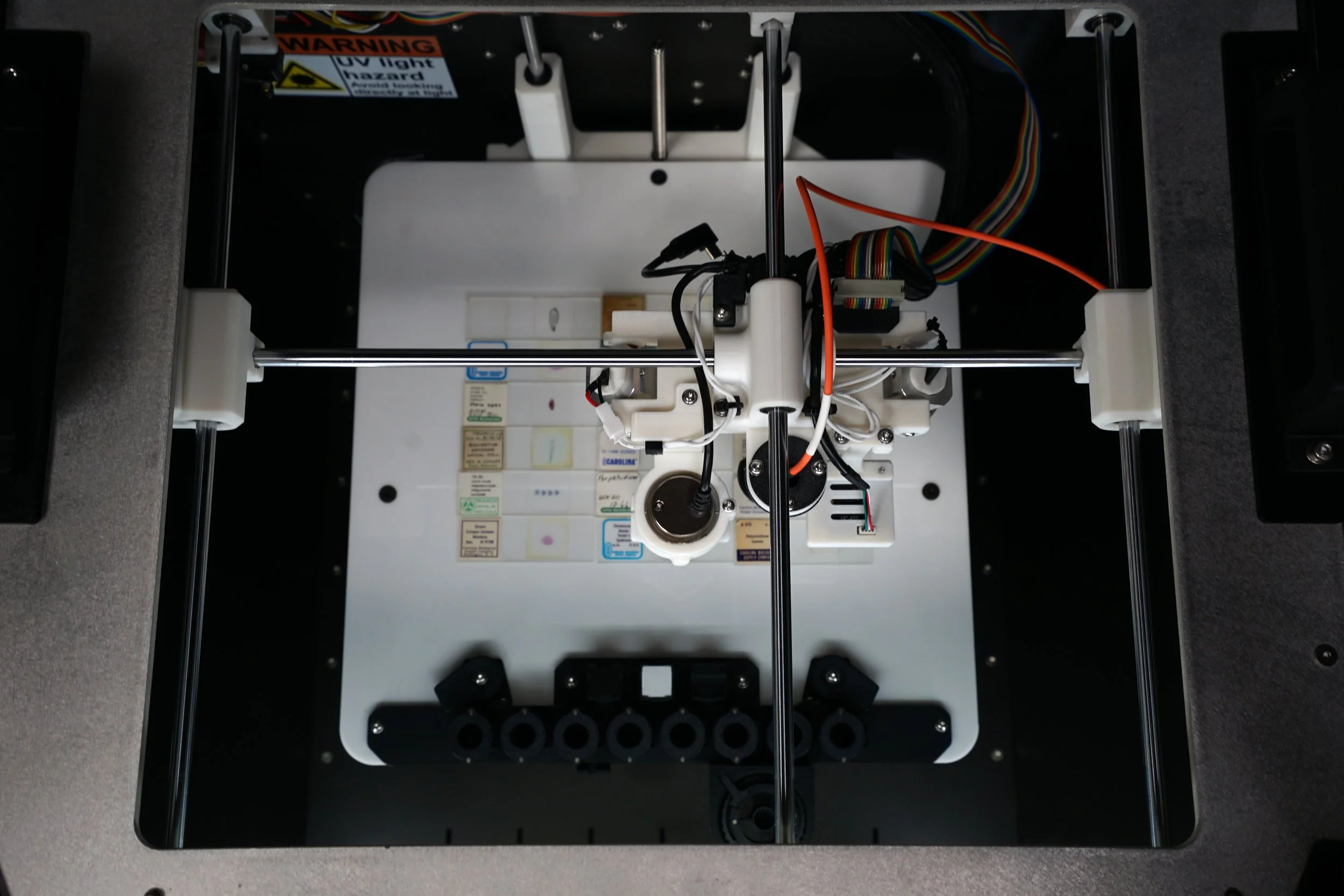
Figure 1—The MODI platform with sensors on a gantry and a large sample volume for high-throughput experimentation.
Beyond Conventional Instrumentation
Many biological analysis workflows rely on either microscopy or well-plate assays. While plate readers provide rapid measurements of fluorescence, absorbance, or luminescence, they typically average results across an entire well and are best suited for liquid samples or homogeneous cell cultures.
As bioengineering moves toward more complex systems - including organoids, engineered tissues, biomaterial scaffolds, and biofilms - important biological processes often occur within localized regions that cannot be captured through bulk measurements alone.
Automated microscopy provides detailed visualization of cellular and tissue structure, while hyperspectral imaging adds chemical information by measuring a complete spectrum at each location. Together, these techniques enable researchers to correlate morphology, composition, and biological activity within the same sample.
Morphological and Chemical Characterization
MODI supports high-resolution imaging of:
Cell cultures
Tissue sections
Biomaterial scaffolds
Organoids
Biofilms
Large-area image stitching and automated acquisition allow researchers to analyze entire samples while preserving microscopic detail.
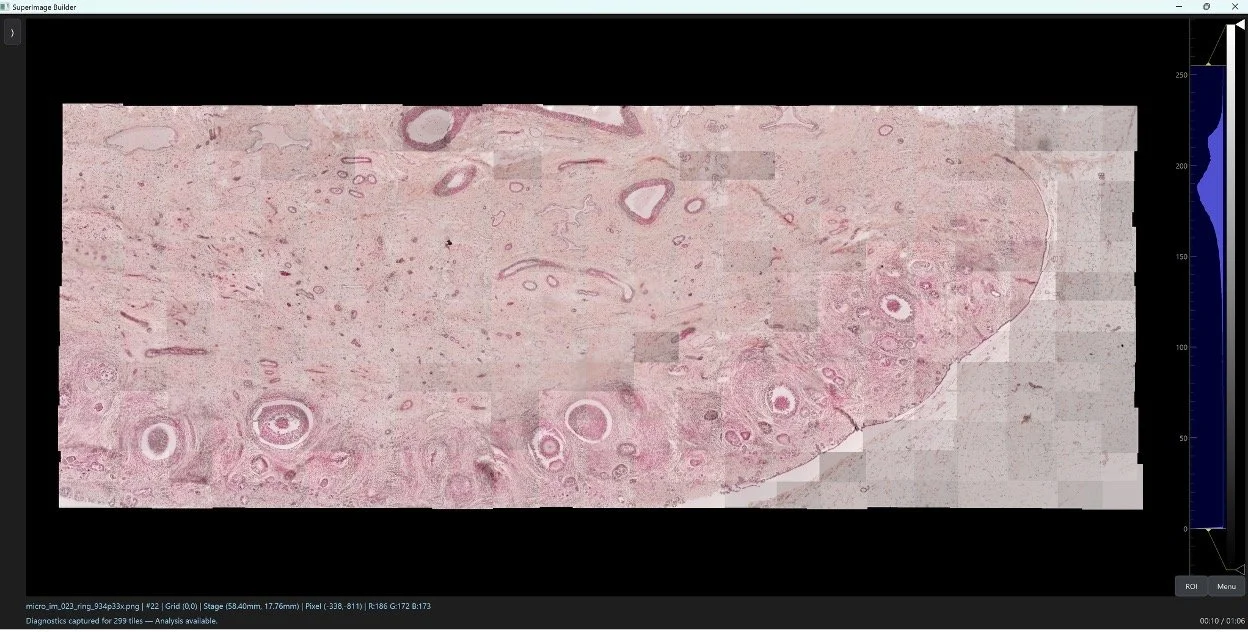
Figure 2—Tissue sample captured with MODI automated microscopy and image stitching.
Point-resolved hyperspectral measurements can reveal subtle differences associated with:
Cellular composition
Tissue heterogeneity
Biomaterial degradation
Hydration changes
Surface contamination
Material interactions
By combining imaging and spectroscopy, researchers gain a more complete understanding of biological systems than either technique can provide independently.
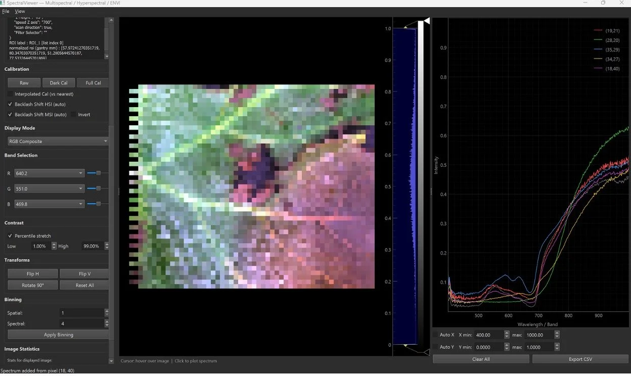
Figure 3—Hyperspectral Image taken with MODI of a small leaf sample and representative spectral signatures of different pigmentations.
Time-Lapse Monitoring of Dynamic Systems
Many biological processes occur over hours, days, or weeks. MODI supports automated time-lapse monitoring by repeatedly imaging and analyzing samples at user-defined intervals.
Applications include:
Cell growth and proliferation
Tissue maturation
Scaffold colonization
Biofilm formation
Wound healing studies
Drug response experiments
Biomaterial degradation
Because measurements can be collected from the same locations over time, researchers can track both structural and chemical changes throughout an experiment.
High-Throughput Bioengineering Workflows
Automated stage positioning and programmable acquisition routines enable systematic analysis across numerous samples, tissue arrays, and biomaterial studies with minimal operator intervention.
By integrating automated microscopy, hyperspectral imaging, and temporal monitoring, MODI provides a versatile platform for studying biological systems that are too spatially complex for conventional bulk measurements and too dynamic for endpoint analysis alone.